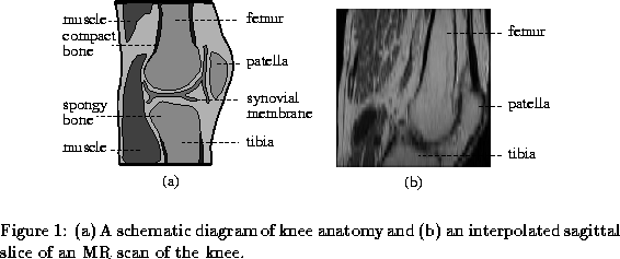


The development of medical, non-invasive imaging techniques such as MR (magnetic resonance) and CT (computed tomography) has created many opportunities for diagnosis, surgical planning and therapy evaluation. Rheumatoid arthritis (RA) evaluation using MR scans of the knee joint is such an application [ 1 ]. It is now possible to monitor the progress of this disease by imaging affected joints and measuring the volume of the inflamed synovial membrane, the primary symptom, shown in figure 1 a. The synovial membrane lines the cavities of the joint and secretes fluid which lubricates and nourishes the cartilage covering the ends of bones; its thickening causes pain and tenderness.
Manually the segmentation and volume calculation is a skilled, laborious and time-consuming task; an automated system would bring benefits by reducing the burden on the radiologist and increasing the scope for evaluation of patients.
Our approach to automating RA monitoring requires the identification of the femur bone which is the largest rigid structure in the image. The femur's boundary can be used as a guide to the location of other anatomy in the knee, such as the synovial membrane. This has proved a significant task to automate with low-level conventional image processing because of poor 3D spatial resolution and artifacts caused by patient movement. A constrained 3D approach to the segmentation is therefore proposed that can model the shape complexity at a global level and can overcome the problems of noise, image artifacts and discontinuous boundaries which are endemic in the knee MR volumes being considered here.
Deformable surfaces offer a compact means of modelling the superficies of a three-dimensional object [ 6 ]. Noise and boundary inconsistencies can be compensated for by the model's connectivity and smoothness properties. The goal of this preliminary work is to produce, reliably and repeatably, a 3D model of the femur from MR volumes of the knee using a deformable surface, based on a discrete geometric mesh [ 8 , 3 ].
However, three specific problems occur when using conventional deformable surfaces with our MR images; identifying the boundary of the feature object, self-intersection of the deformable surface reported in [ 8 ] and leakage of the deformable surface out of broken or weakly defined object boundaries as noted in [ 7 ]. These problems are addressed by rejecting intensity gradients as a means of identifying edges in favour of the novel approach of probabilistic labelling driving the deformation process. We also introduce a relaxation phase during the deformation stage to prevent self-intersection and leakage, by identifying possible surface errors and contracting the local surface in response to them.


N Hill