Medical images are often complex, of poor visual quality and open to subjective interpretation. Machine vision can be used to help analyse images, such as radiographs and magnetic resonance (MR) scans, leading to more effective use of an expert's time.
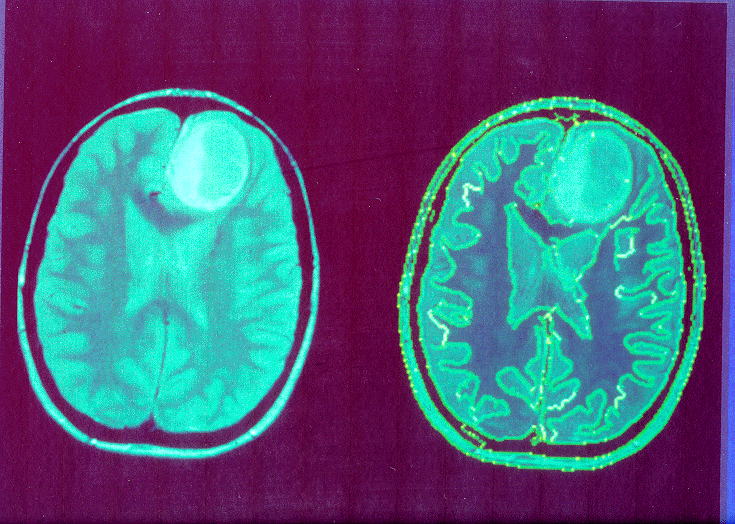
When planning radiation therapy of brain tumours from MR scans of the
head, it is necessary to apply to most effective dose of radiation to
the tumour, yet cause least damage to the surrounding tissue. This is a
complex task demanding 3D visualization. An interactive system is being
developed to produce 3D representations of the head, by combining the
information from a series of cross-sectional images. Segmentation
techniques are used to semi-automatically identify the tumour and other
anatomical structures. One approach is for the clinician to select a
point within an area of interest, such as the tumour. The system then
locates the surrounding points which possess similar characteristics,
within a given range of variability. This is repeated for other regions,
and other images, to produce a 3D model. Given this model, an optimal
treatment plan can be generated.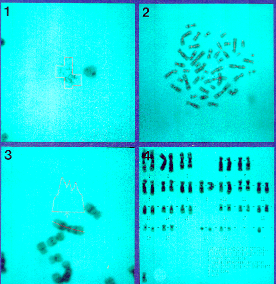 Chromosone analysis (CA) for the diagnosis of genetic disorders is
usually performed manually. The efficiency of the process can be greatly
enhanced by interactive machine vision. An appropriate cell is
identified using textural analysis of the microscopic images. The axes
of symmetry of the chromosomes are found, chromosome length measured,
and the constricted region (centromere) located from lateral profiles.
The pattern of stain uptake is then measured and a characteristic
profile obtained. The identity of the chromosome is determined so it can
be placed in the appropriate position in an ordered array. Long
chromosomes may form complex overlaps which can be resolved using
geometric evidence. As the system is intereactive, misidentification or
failure to identify chromosomes can be corrected by the user. This
approach to CA has been shown to increase laboratory throughput by a
factor of two, thus allowing more widespread use.
Chromosone analysis (CA) for the diagnosis of genetic disorders is
usually performed manually. The efficiency of the process can be greatly
enhanced by interactive machine vision. An appropriate cell is
identified using textural analysis of the microscopic images. The axes
of symmetry of the chromosomes are found, chromosome length measured,
and the constricted region (centromere) located from lateral profiles.
The pattern of stain uptake is then measured and a characteristic
profile obtained. The identity of the chromosome is determined so it can
be placed in the appropriate position in an ordered array. Long
chromosomes may form complex overlaps which can be resolved using
geometric evidence. As the system is intereactive, misidentification or
failure to identify chromosomes can be corrected by the user. This
approach to CA has been shown to increase laboratory throughput by a
factor of two, thus allowing more widespread use.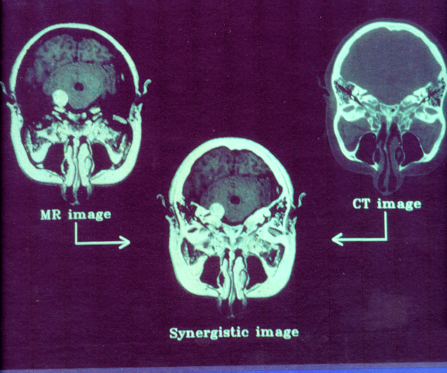 Different types of medical image yield different information. For
example, MR scans provide soft tissue information, yet nothing regarding
bones. The converse is true of computed tomography (CT) scans. When
planning surgery, information from both CT and MR scans may be needed.
Machine vision techniques can be used to collate information from the
different scans, thus producing a single 3D image showing the
relationship between single 3D image showing the relationship between
important features. This has been achieved, for example, by
interactively labelling 12-16 matching features in the original images.
Bone is identified in the CT scan by locating areas with grey levels
above a certain threshold value. Identifying tissue structures in the MR
scan requires more sophisticated segmentation techniques. This process
is now being developed to use known anatomical relationships to
automatically match 3D models derived from different medical images.
Different types of medical image yield different information. For
example, MR scans provide soft tissue information, yet nothing regarding
bones. The converse is true of computed tomography (CT) scans. When
planning surgery, information from both CT and MR scans may be needed.
Machine vision techniques can be used to collate information from the
different scans, thus producing a single 3D image showing the
relationship between single 3D image showing the relationship between
important features. This has been achieved, for example, by
interactively labelling 12-16 matching features in the original images.
Bone is identified in the CT scan by locating areas with grey levels
above a certain threshold value. Identifying tissue structures in the MR
scan requires more sophisticated segmentation techniques. This process
is now being developed to use known anatomical relationships to
automatically match 3D models derived from different medical images.